
What is ankle impingement?
This is a common cause of pain at the front of the ankle joint. ‘Impingement’ describes the presence of excess bone or soft tissue within the ankle that causes bumpering shin bone and ankle bone. This occurs when walking and can decrease the range of up - down ankle movement. Excess bone spurs can often be seen on xrays along the front of the ankle joint. This pattern is classically referred to as ‘footballer’s ankle’. Impingement can also occur from soft tissue. This is typically scar tissue that forms from injury such as ankle sprains, fractures or from sports. It has an appearance and texture similar to cotton wool when seen with the naked eye. Like bone impingement, this tissue can be caught in the front of the ankle joint as it moves and cause bumpering.
What are the symptoms?
Impingement causes pain at the front of the ankle, as well as to the outer aspect. It is often worse on standing, or when trying to bring the ankle upwards. The pain is often described as a dull aching pain that can stop walking over long distances, as well as sports activity. In the case of the ‘anterolateral’ soft tissue impingement, the location of the pain is discrete, and can be associated with a fullness or swelling. Pressing on the outer aspect of the ankle joint and bringing the foot upwards will usually recreate the symptoms. In some cases, the ankle may feel loose and that it rolls over. These particular symptoms indicates ankle instability from prior ligament injury, which must also be addressed.
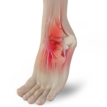
The picture above demonstrates where the pain from impingement is typically felt. This tends to be along the front and outer aspect of the ankle joint
Assessment
Your symptoms and a thorough physical examination will determine the type of treatment that will be most appropriate for you.
Xrays will help to determine the condition of the ankle joint and detect bone spurs along the shin and ankle bone, as shown below. Further imaging such as a MRI can be used to investigate the soft tissue around the ankle joint, highlighting the presence of scar tissue, cartilage damage and ligament injury. Inflammation of the tissues, abnormal fluid accumulation and areas of bone stress reaction may also be seen
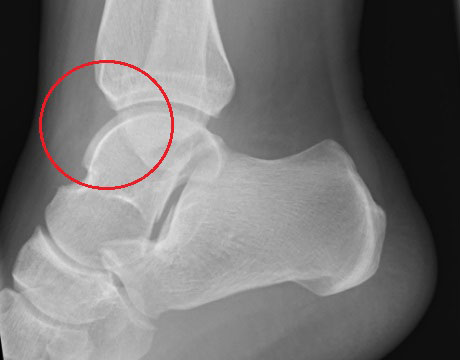
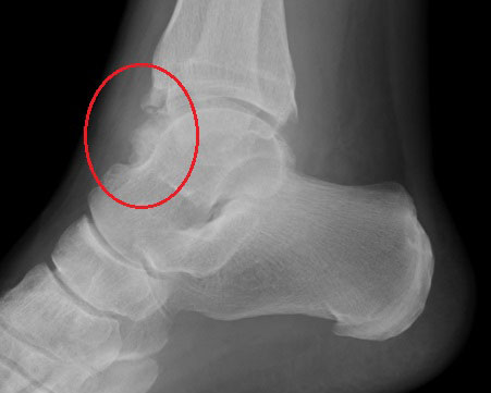
The first xray above shows a normal ankle. The red circle highlights the front of the ankle. Here there is no bone impingement. The second xray shows an accumulation of bone along the front of the anke joint. This causes a ‘bumper’ effect as the joint moves up and down.
What treatment is available?
Non surgical
Early management options are outlined below. These may improve your symptoms, but will not address the underlying bone or soft tissue cause of anterior ankle impingement.
- Calf stretching exercise.
As the ankle stiffens with impingement, the Achilles tendon may begin to shorten and contract. Regular Achilles tendon exercise will help to increase the range of ankle movement, particularly upwards.
- Heel inserts and pad
Using a shoe with an elevated heel, or a heel lift inside the shoe. This means the foot and ankle do not need to come upward towards the shin bone when walking. Therefore less impingement and bumpering occurs.
- Activity modification
Avoiding certain activities will help alleviate symptoms. Examples include walking uphill or playing sports.
- Ankle brace
A supportive ankle brace will prevent excessive ankle movement and hence irritation within the joint. This will provide short term symptom relief, but is not a long term solution.
- Injection of local anaesthetic and steroid
This can be safely delivered into the joint in the clinic or with ultrasound guidance. The local anaesthetic will switch off the pain in the first few days after the injection, whilst the steroid will take several weeks to dampen inflammation in the joint. In a small percentage of patients, the injection can cause a symptom flare for a few days. Further information can be found here.
Surgery
The aim of surgery is to remove the bone spur or soft tissue causing the impingement. It should be considered once the non surgical options described above have been attempted.
Key hole surgery
The operation of choice is normally key hole surgery (called ‘ankle arthroscopy’) to look inside the ankle. This is achieved through two small incisions in front of the ankle. The joint is gradually inflated with water. It can be thoroughly visualised and small instruments and shavers can be used to remove the impinging tissue to clean out the joint. The further advantage of this procedure is that Mr Davda can also investigate for other causes of pain such as damaged cartilage or early arthritis.
The results of surgery for ankle surgery are very good with about an 80-90% improvement in symptoms. The ankle will feel sore for a few weeks after surgery and the swelling from the procedure may take several months to settle. Physiotherapy can begin approximately two weeks following the operation, once the wounds have been checked. Ankle arthroscopy is described in greater detail here.
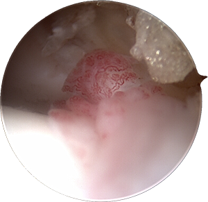
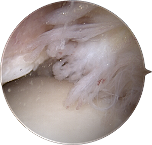
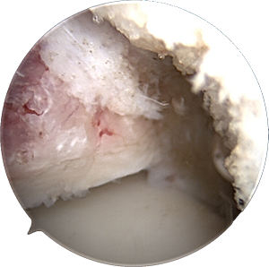
The photos above are taken during ankle arthroscopy. The first picture shows inflammed red looking tissue that is often a cause of pain. The second picture shows the ‘fluffy’ cotton wool like scar tissue that has developed over time and causes pain and bumpering between the joint surfaces. A small shaver measuring a few millimetres is used to clear the scar tissue as shown in the picture.
Calf release
In some cases, the calf may have tightened over the course of time and it may be difficult to bring the ankle fully upwards. The calf muscle may need a small procedure to release it and is described in greater detail in ‘Calf tightness’.